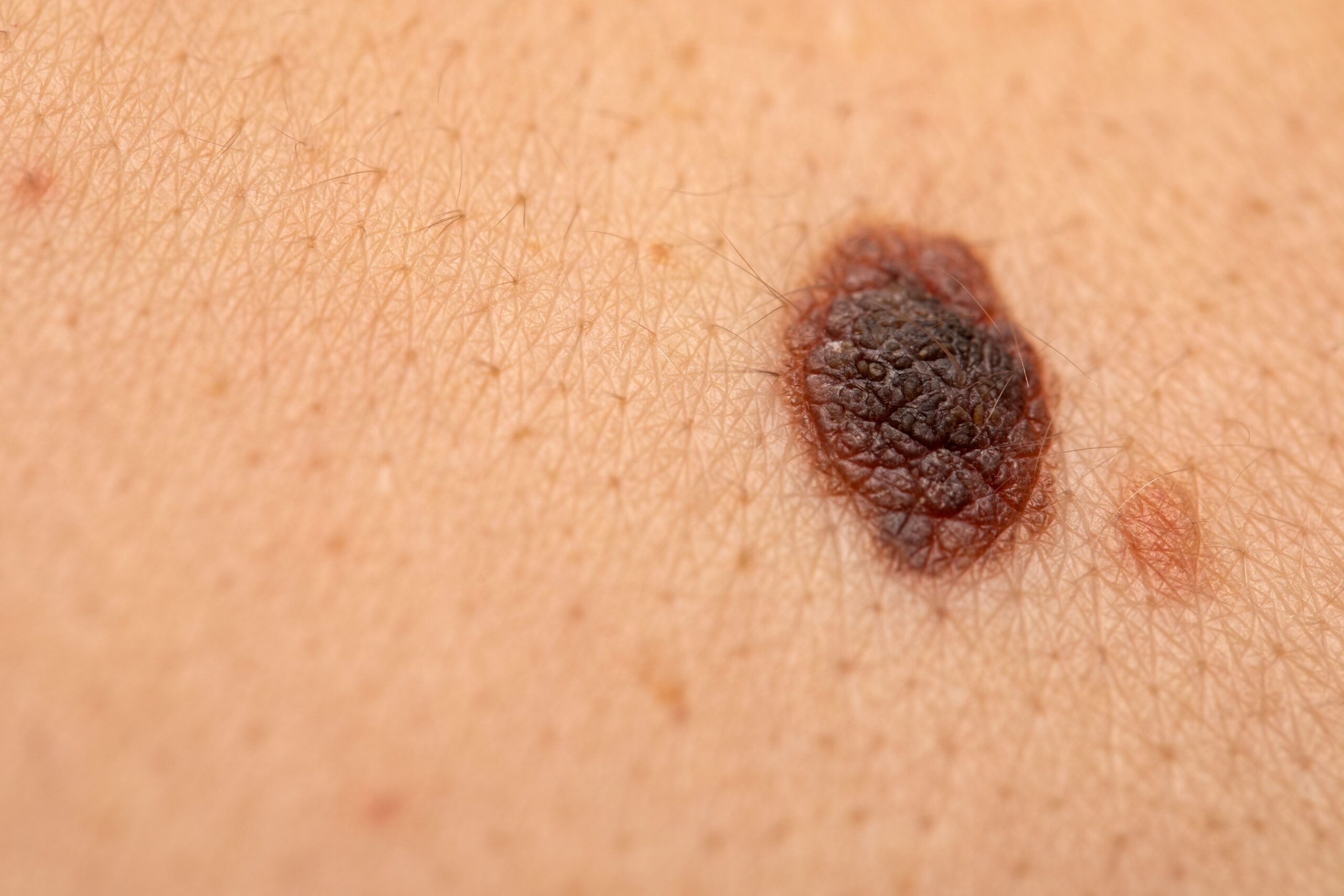
As the incidence of melanoma and biopsies performed have increased, death rates have remained relatively stagnant. Although previous explanations often cite the role of different diagnostic thresholds among pathologists, overdiagnosis, or advances in melanoma treatment, a study recently published in British Journal of Dermatology suggests that further research is needed to determine any causal relationships in these trends.1
Skin injury | Image credit: Ocskay Mark – stock.adobe.com
According to a 2019 Global Burden of Disease Study, there was an average global increase of 1.13% in cases of cutaneous melanoma (95% CI, 0.93-1.32) each year between 1990 and 2019. During this period, age-standardized mortality rates decreased by 0.27% (95% CI, 0.19-0.36).2 As the authors of the present study point out, this trend is not fully understood. Analysis of behavioral patterns leading to increased sun exposure (use of tanning beds, vacation destinations, etc.), changes in melanoma treatment interventions and diagnostic approaches (lowered diagnostic thresholds, overdiagnosis, etc.) have not offered adequate explanations for this discrepancy.
To address this knowledge gap, Nielsen et al conducted a study to evaluate the various factors associated with melanoma, including treatment, ultraviolet radiation. [UV] exposure, diagnosis patterns and more that could affect incidence and mortality rates in Denmark.
Data were collected from a national health system to survey 5.5 million people. Biopsies from patients with melanocytic skin lesions from the Danish Pathology Data Bank (DPDB) between 1999 and 2019 were retrospectively analyzed to investigate subsequent diagnoses and mortalities in this population. Among the surveyed patients, the authors created 3 age groups: 15-44 years; 45-59 years; and 60 years or older.1
Among the 5.5 million people, melanocyte-related diagnoses were seen in 1,434,798 biopsies. Of these biopsies, an overwhelming 93.1% (n = 1,336,361) were benign, while 3.5% (n = 49,895) were invasive melanoma, 2.2% (n = 31,127) were considered atypical and 1.2% (n = 17,415). site melanoma. There were 11,572 people who experienced more than 1 melanoma diagnosis during the study period, and the average age at biopsy fell between 37 and 40 years. In addition, 57.5% (n = 405,098) of people had only a single biopsy, compared with 42.5% (n = 299,584) who underwent multiple.
The researchers observed that biopsy rates in men grew by 153% (from 540 to 1364 per 100,000 population per year) between 1999 and 2011, and rates in women grew by 118% (from 1272 to 2772). In 2019, these rates had dropped in men and women by 20% (1364 to 1085) and 22% (2772 to 2165), respectively. In addition, the authors noted that younger individuals and women in general underwent more biopsies compared to older individuals and men.
Cases of benign lesions in men and women grew by 149% (504 to 1256) and 114% (1227 to 2631) between 1999 and 2011. With a similar trend to biopsy rates, reports of these cases fall in men and women by 25% (1256 to 937) and 24% (2631 to 1991), respectively. Regarding atypical skin lesions, the prevalence of these cases increased by 1928% for men (3 to 61 per 100,000 per year) and by 1686% (4 to 77) for women over the entire period of study In addition, between 1999 and 2019, the incidence of melanoma in situ increased by 476% (5.12 to 29.48) in men and by 357% (7.26 to 33.21). The prevalence of invasive melanoma grew by 87% in both men and women (28 in 1999; 34 to 63, respectively) between 1999 and 2011. In the following 8 years, each group experienced a 9% increase in incidence .
Over the 21-year study period, the authors wrote, death rates ranged from 4.57 to 6.79 in men and 3.98 to 5.01 in women. During this time, the group of 45-60 years presented a similar pattern; however, the incidences were estimated to be 3 times higher compared to the younger age group. Although the older age group also had a similar trend, the incidence among this group was 8 times higher than that of the younger age group.
The authors wrote that their analysis revealed a strong association between biopsy rates and benign lesions (R = 0.99), as well as melanoma (R = 0.82 in men; 0.86 in women), atypical (R = 0.69) and in situ (R = 0.67) incidence. A linear regression analysis further showed that for each additional biopsy, the incidence of melanoma would increase by 0.01 (95% CI 0.01–0.02 in men and 0.00–0.01 in women) .
We were unable to support increased UV exposure as an explanation for the increased incidence of melanoma before or during the study period. Rather, exposure is expected to decrease after several public information campaigns, the authors concluded.
The other explanations seem more plausible. The treatment of late-stage melanoma has gradually improved since 2010. The data show an increase in diagnostic activity, and especially in the population groups with the lowest risk of melanoma. The findings are consistent with overdiagnosis, but earlier diagnosis and treatment may at least partially explain the findings. The rapid increase in atypical and in situ melanoma relative to invasive melanoma could be associated with changes in pathologists’ threshold for specifying these diagnoses. It is conceivable that the threshold for atypical and in situ melanoma, as well as for invasive melanoma, has decreased due to increased awareness of melanoma.
References
1. Nielsen JB, Kristiansen IS, Thapa S. Increasing incidence of melanoma with unchanged mortality: more alone, better treatment, increased diagnostic activity, overdiagnosis or lowered diagnostic threshold? Br J Dermatol. 202:ljae175. doi:10.1093/bjd/ljae175
2. Li Z, Fang Y, Chen H, et al. Spatiotemporal trends in the global burden of melanoma in 204 countries and territories from 1990 to 2019: results from the 2019 Global Burden of Disease Study. neoplasia. 2022;24(1):12-21. doi:10.1016/j.neo.2021.11.013
#Exploring #melanoma #incidence #mortality #rates #calls #research
Image Source : www.ajmc.com
